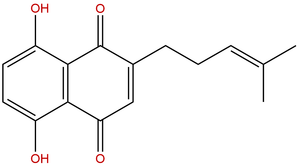
DeoxyshikoninCAS No.:43043-74-9
|
||||||||||
 |
|
|
||||||||

| Catalogue No.: | BP3119 |
| Formula: | C16H16O4 |
| Mol Weight: | 272.3 |
Product name: Deoxyshikonin
Synonym name:
Catalogue No.: BP3119
Cas No.: 43043-74-9
Formula: C16H16O4
Mol Weight: 272.3
Botanical Source:
Physical Description: Powder
Type of Compound: Quinones
Purity: 95%~99%
Analysis Method: HPLC-DAD or/and HPLC-ELSD
Identification Method: Mass, NMR
Packing: Brown vial or HDPE plastic bottle
The product could be supplied from milligrams to grams. Inquire for bulk scale.
We provide solution to improve the water-solubility of compounds, thereby facilitating the variety of activity tests and clinic uses.
For Reference Standard and R&D, Not for Human Use Directly.
Description:
Deoxyshikonin and dodecyl gallate show significantly synergic antimicrobial activity with penicillin in vivo and in vitro, and can effectively reduce nasopharyngeal and lung colonization caused by different penicillin-resistant pneumococcal serotypes. Deoxyshikonin exerts very good radical scavenging activities toward ABTS+ but shows moderate inhibition of DPPH·, and shows cytotoxic activities. Deoxyshikonin may be a new drug candidate for wound healing and treatment of lymphatic diseases.
References:
Evid Based Complement Alternat Med. 2013;2013:148297.
Enhancement of Lymphangiogenesis In Vitro via the Regulations of HIF-1α Expression and Nuclear Translocation by Deoxyshikonin.
The objectives of this study were to determine the effects of Deoxyshikonin on lymphangiogenesis.
METHODS AND RESULTS:
Deoxyshikonin enhanced the ability of human dermal lymphatic microvascular endothelial cells (HMVEC-dLy) to undergo time-dependent in vitro cord formation. Interestingly, an opposite result was observed in cells treated with shikonin. The increased cord formation ability following Deoxyshikonin treatment correlated with increased VEGF-C mRNA expression to higher levels than seen for VEGF-A and VEGF-D mRNA expression. We also found that Deoxyshikonin regulated cord formation of HMVEC-dLy by increasing the HIF-1 α mRNA level, HIF-1 α protein level, and the accumulation of HIF-1 α in the nucleus. Knockdown of the HIF-1 α gene by transfection with siHIF-1 α decreased VEGF-C mRNA expression and cord formation ability in HMVEC-dLy. Deoxyshikonin treatment could not recover VEGF-C mRNA expression and cord formation ability in HIF-1 α knockdown cells. This indicated that Deoxyshikonin induction of VEGF-C mRNA expression and cord formation in HMVEC-dLy on Matrigel occurred mainly via HIF-1 α regulation. We also found that Deoxyshikonin promoted wound healing in vitro by the induction of HMVEC-dLy migration into the wound gap. This study describes a new effect of Deoxyshikonin, namely, the promotion of cord formation by human endothelial cells via the regulation of HIF-1 α .
CONCLUSIONS:
The findings suggest that Deoxyshikonin may be a new drug candidate for wound healing and treatment of lymphatic diseases.
Front Microbiol. 2015 May 20;6:479.
Antibacterial effects of Traditional Chinese Medicine monomers against Streptococcus pneumoniae via inhibiting pneumococcal histidine kinase (VicK).
Two-component systems (TCSs) have the potential to be an effective target of the antimicrobials, and thus received much attention in recent years. VicK/VicR is one of TCSs in Streptococcus pneumoniae (S. pneumoniae), which is essential for pneumococcal survival. We have previously obtained several Traditional Chinese Medicine monomers using a computer-based screening.
METHODS AND RESULTS:
In this study, either alone or in combination with penicillin, their antimicrobial activities were evaluated based on in vivo and in vitro assays. The results showed that the MICs of 5'-(Methylthio)-5'-deoxyadenosine, octanal 2, 4-dinitrophenylhydrazone, Deoxyshikonin, kavahin, and dodecyl gallate against S. pneumoniae were 37.1, 38.5, 17, 68.5, and 21 μg/mL, respectively. Time-killing assays showed that these compounds elicited bactericidal effects against S. pneumoniae D39 strain, which led to a 6-log reduction in CFU after exposure to compounds at four times of the MIC for 24 h. The five compounds inhibited the growth of Streptococcus pyogenes, Streptococcus mitis, Streptococcus mutans or Streptococcus pseudopneumoniae, meanwhile, Deoxyshikonin and dodecyl gallate displayed strong inhibitory activities against Staphylococcus aureus. These compounds showed no obvious cytotoxicity effects on Vero cells. Survival time of the mice infected by S. pneumoniae strains was prolonged by the treatment with the compounds. Importantly, all of the five compounds exerted antimicrobial effects against multidrug-resistant clinical strains of S. pneumoniae. Moreover, even at sub-MIC concentration, they inhibited cell division and biofilm formation. The five compounds all have enhancement effect on penicillin. Deoxyshikonin and dodecyl gallate showed significantly synergic antimicrobial activity with penicillin in vivo and in vitro, and effectively reduced nasopharyngeal and lung colonization caused by different penicillin-resistant pneumococcal serotypes. In addition, the two compounds also showed synergic antimicrobial activity with erythromycin and tetracycline.
CONCLUSIONS:
Taken together, our results suggest that these novel VicK inhibitors may be promising compounds against the pneumococcus, including penicillin-resistant strains.
Food Chemistry, 2008 , 106 (1) :2-10.
Antioxidants from a Chinese medicinal herb - Lithospermum erythrorhizon
Seven compounds, Deoxyshikonin (1), β,β-dimethylacrylshikonin (2), isobutylshikonin (3), shikonin (4), 5,8-dihydroxy-2-(1-methoxy-4-methyl-3-pentenyl)-1,4-naphthalenedione (5), β-sitosterol (6) and a mixture of two caffeic acid esters [7 (7a,7b)] were isolated from Lithospermum erythrorhizon Sieb et. Zucc. and identified by spectroscopic methods. Among them, compound 5 was isolated from this plant species for the first time.
METHODS AND RESULTS:
The antioxidant activities of the seven compounds were compared and evaluated through Rancimat method, reducing power and radical scavenging activity. Results showed that, except compound 6, another 6 compounds all exhibited obvious antioxidant activities against four different methods. Compounds 4 and 7 exerted much more potent antioxidant effects on retarding the lard oxidation than that of BHT and both were found to exhibit strong reducing power. Their antioxidant activities, assessed by Rancimat method and reducing power, decreased in the following order, respectively: compound 7 > 4 > BHT > 2 > 3 > 5 > 1 > 6 and compound 7 > BHT > 4 > 2 approximately 3 approximately 5 > 1> 6. In addition, compounds 1-5 all exerted very good radical scavenging activities toward ABTS+ but showed moderate inhibition of DPPH·, while compound 7 presented as a powerful radical scavenger against both ABTS·+ and DPPH·.
CONCLUSIONS:
Thus, our results suggested that L. erythrorhizon could be a promising rich source of natural antioxidants.