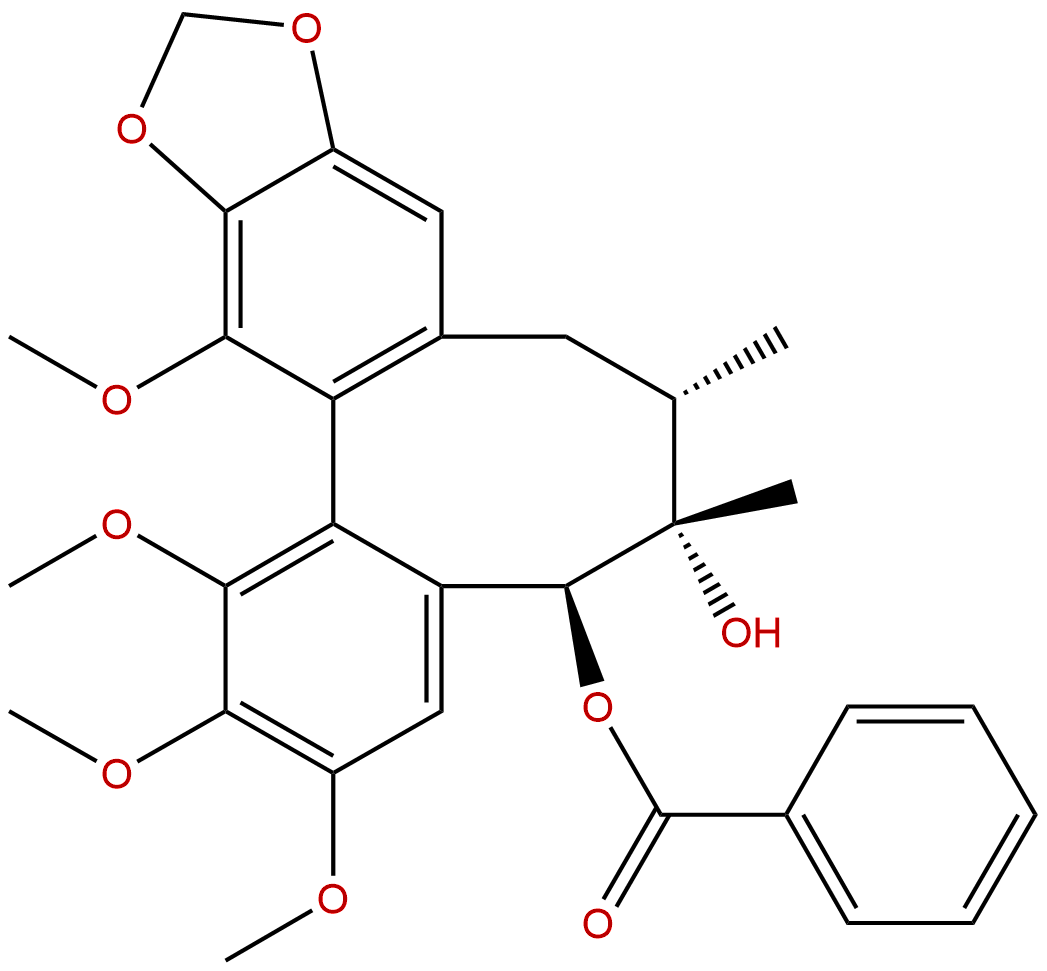
Schizantherin ACAS No.:58546-56-8
|
||||||||||
 |
|
|
||||||||

| Catalogue No.: | BP1267 |
| Formula: | C30H32O9 |
| Mol Weight: | 536.577 |
Product name: Schizantherin A
Synonym name: Gomisin C; Schisantherin A; Wuweizi ester A
Catalogue No.: BP1267
Cas No.: 58546-56-8
Formula: C30H32O9
Mol Weight: 536.577
Botanical Source: Schisandra chinensis(Turcz.)Baill.
Physical Description:
Type of Compound: Lignans
Purity: 95%~99%
Analysis Method: HPLC-DAD or/and HPLC-ELSD
Identification Method: Mass, NMR
Packing: Brown vial or HDPE plastic bottle
Storage: Store in a well closed container, protected from air and light. Put into refrigerate or freeze for long term storage.
Whenever possible, you should prepare and use solutions on the same day. However, if you need to make up stock solutions in advance, we recommend that you store the solution as aliquots in tightly sealed vials at -20℃. Generally, these will be useable for up to two weeks.
The product could be supplied from milligrams to grams
Inquire for bulk scale.
Description:
Schisantherin A exhibits anti-tussive, sedative, anti-inflammatory, antioxidant, anti-osteoporotic, neuroprotective, cognition enhancing, and cardioprotective activities. Schisantherin A can significantly attenuate Aβ1-42-induced learning and memory impairment and noticeably improve the histopathological changes in the hippocampus. Schisantherin A exhibits neuroprotection against 1-methyl-4-phenylpyridinium ion (MPP(+)) through the regulation of two distinct pathways including increasing CREB-mediated Bcl-2 expression and activating PI3K/Akt survival signaling.
References:
PLoS One. 2013 Apr 19;8(4):e61590.
Protective role of deoxyschizandrin and schisantherin A against myocardial ischemia-reperfusion injury in rats.
METHODS AND RESULTS:
Anesthetized male rats were treated once with deoxyschizandrin(DSD) and Schisantherin A(STA) (each 40 μmol/kg) through the tail vein after 45 min of ischemia, followed by 2-h reperfusion. Cardiac function, infarct size, biochemical markers, histopathology and apoptosis were measured and mRNA expression of gp91 (phox) in myocardial tissue assessed by RT-PCR. Neonatal rat cardiomyocytes were pretreated with DSD and STA and then damaged by H2O2. Cell apoptosis was tested by a flow cytometric assay. Compared with the I/R group: (i) DSD and STA could significantly reduce the abnormalities of LVSP, LVEDP, ±dp/dtmax and arrhythmias, thereby showing their protective roles in cardiac function; (ii) DSD and STA could significantly attenuate the infarct size and MDA release while increasing SOD activity, suggesting a role in reducing myocardial injury; (iii) tissue morphology and myocardial textual analysis revealed that DSD and STA mitigated changes in myocardial histopathology; (iv) DSD and STA decreased apoptosis (33.56±2.58% to 10.28±2.80% and 10.98±1.99%, respectively) and caspase-3 activity in the myocardium (0.62±0.02 OD/mg to 0.38±0.02 OD/mg and 0.32±0.02 OD/mg, respectively), showing their protective effects upon cardiomyocytes; and (v) DSD and STA had similar protective effects on I/R injury as those seen with the positive control metoprolol. In vitro, DSD and STA could significantly decrease the apoptosis of neonatal cardiomyocytes.
CONCLUSIONS:
These data suggest that DSD and STA can protect against myocardial I/R injury. The underlining mechanism may be related to their role in inhibiting cardiomyocyte apoptosis.
Biochem Biophys Res Commun. 2014 Jul 4;449(3):344-50.
Schisantherin A suppresses osteoclast formation and wear particle-induced osteolysis via modulating RANKL signaling pathways.
Receptor activator of NF-κB ligand (RANKL) plays critical role in osteoclastogenesis. Targeting RANKL signaling pathways has been a promising strategy for treating osteoclast related bone diseases such as osteoporosis and aseptic prosthetic loosening. Schisantherin A (SA), a dibenzocyclooctadiene lignan isolated from the fruit of Schisandra sphenanthera, has been used as an antitussive, tonic, and sedative agent, but its effect on osteoclasts has been hitherto unknown.
METHODS AND RESULTS:
In the present study, SA was found to inhibit RANKL-induced osteoclast formation and bone resorption. The osteoclastic specific marker genes induced by RANKL including c-Src, SA inhibited OSCAR, cathepsin K and TRAP in a dose dependent manner. Further signal transduction studies revealed that SA down-regulate RANKL-induced nuclear factor-kappaB (NF-κB) signaling activation by suppressing the phosphorylation and degradation of IκBα, and subsequently preventing the NF-κB transcriptional activity. Moreover, SA also decreased the RANKL-induced MAPKs signaling pathway, including JNK and ERK1/2 posphorylation while had no obvious effects on p38 activation. Finally, SA suppressed the NF-κB and MAPKs subsequent gene expression of NFATc1 and c-Fos. In vivo studies, SA inhibited osteoclast function and exhibited bone protection effect in wear-particle-induced bone erosion model. Taken together, SA could attenuate osteoclast formation and wear particle-induced osteolysis by mediating RANKL signaling pathways.
CONCLUSIONS:
These data indicated that SA is a promising therapeutic natural compound for the treatment of osteoclast-related prosthesis loosening.
HPLC of Schizantherin A
