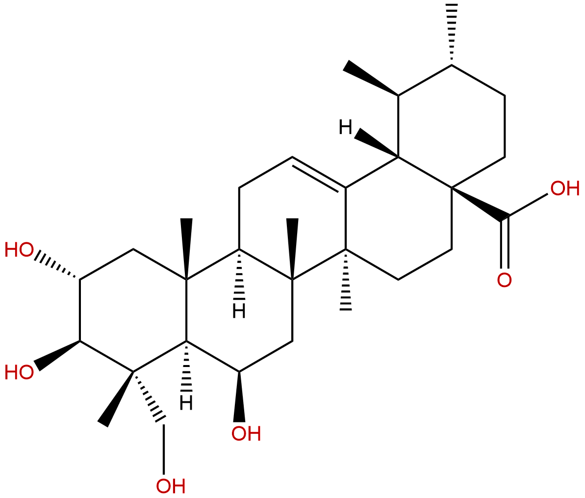
Madecassic acidCAS No.:18449-41-7
|
||||||||||
 |
|
|
||||||||

| Catalogue No.: | BP0910 |
| Formula: | C30H48O6 |
| Mol Weight: | 504.708 |
Product name: Madecassic acid
Synonym name: Brahmic acid; 6-Hydroxyasiatic acid
Catalogue No.: BP0910
Cas No.: 18449-41-7
Formula: C30H48O6
Mol Weight: 504.708
Botanical Source: Centella asiatica (L.)Urb.
Physical Description:
Type of Compound: Triterpenoids
Purity: 95%~99%
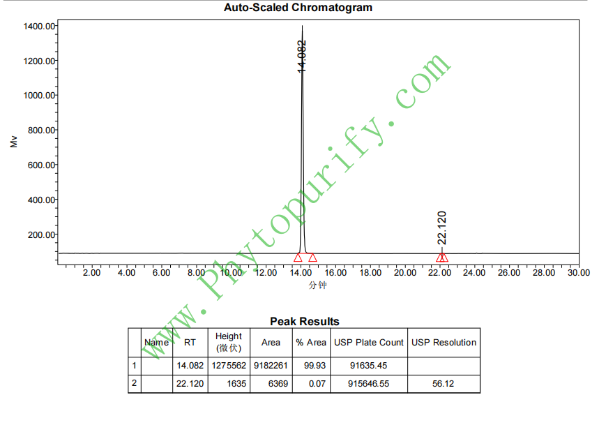
Analysis Method: HPLC-DAD or/and HPLC-ELSD
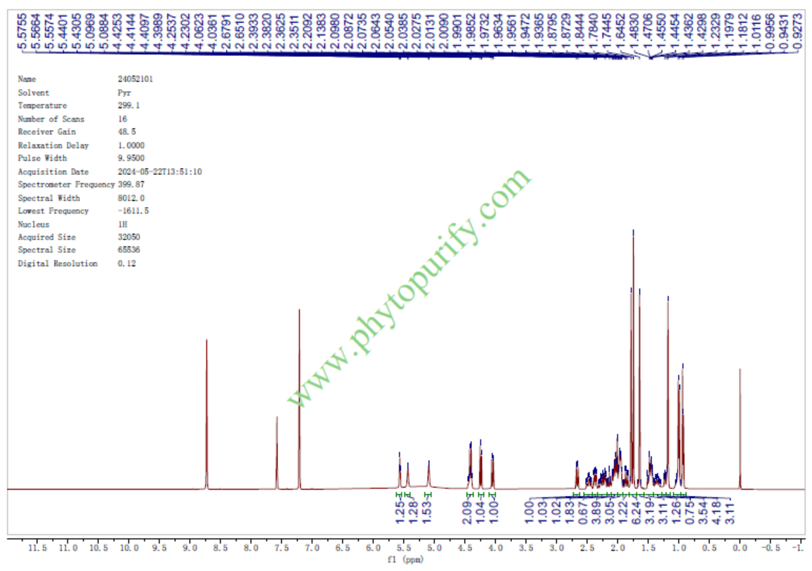
Identification Method: Mass, NMR
Packing: Brown vial or HDPE plastic bottle
Storage: Store in a well closed container, protected from air and light. Put into refrigerate or freeze for long term storage.
The product could be supplied from milligrams to grams
Inquire for bulk scale.
Description:
Madecassic acid has anti-diabetic, anti- tumor, wound-healing, and anti-inflammatory properties, it can improve glycemic control and hemostatic imbalance, lower lipid accumulation, and attenuate oxidative and inflammatory stress in diabetic mice. It can protect against hypoxia-induced oxidative stress in retinal microvascular endothelial cells via ROS-mediated endoplasmic reticulum stress. It inhibited the esspession of NOS, COX-2, TNF-alpha, IL-1beta, IL-6, and the downregulation of NF-kappaB activation.
References:
Biomed Pharmacother. 2016 Dec;84:845-852.
Madecassic Acid protects against hypoxia-induced oxidative stress in retinal microvascular endothelial cells via ROS-mediated endoplasmic reticulum stress.
Madecassic acid (MA) is an abundant triterpenoid in Centella asiatica (L.) Urban. (Apiaceae) that has been used as a wound-healing, anti-inflammatory and anti-cancer agent. Up to now, the effects of MA against oxidative stress remain unclear.
METHODS AND RESULTS:
In this study, we investigated the effect of MA and its mechanisms on hypoxia-induced human Retinal Microvascular Endothelial Cells (hRMECs). hRMECs were pre-treated with different concentrations of MA (0-50μM) for 30min before being incubated under hypoxia condition (37°C, 5% CO2 and 95% N2). Cell apoptosis was evaluated with MTT assay and TUNEL staining, and the expression of apoptosis- and endoplasmic reticulum (ER) stress-related molecules was assessed with western blotting and RT-PCR analysis. Intracellular ROS level was evaluated using DCFH-DA. Intracellular malondialdehyde (MDA), dehydrogenase (LDH), glutathione peroxidase (GSH-PX) and superoxide dismutase (SOD) were evaluated using related Kits. Activating transcription factor 4 (ATF4) nuclear translocation was assessed with western blotting analysis and immunofluorescence staining. MA significantly reduced oxidative stress in hypoxia-induced hRMECs, as shown by increased cell viability, SOD and GSH-PX leakage, decreased TUNEL- and ROS-positive cell ratio, LDH and MDA leakage, caspase-3 and -9 activity, and Bax/Bcl-2 ratio. In addition, MA also attenuated hypoxia-induced ER stress in hRMECs, as shown by reduced mRNA levels of glucose-regulated protein 78 (GRP78), C/EBP homologous transcription factor (CHOP), protein levels of cleaved activating transcription factor 6 (ATF6) and inositol-requiring kinase/endonuclease 1 alpha (IRE1α), phosphorylation of pancreatic ER stress kinase (PERK) and eukaryotic initiation factor 2 alpha (eIF2α), cleaved caspase-12 and ATF4 translocation to nucleus.
CONCLUSIONS:
The current study indicated that the regulation of oxidative stress and ER stress by MA would be a promising therapy to reverse the process and development of hypoxia-induced hRMECs dysfunction.
Nutrients. 2015 Dec 2;7(12):10065-75.
Anti-Diabetic Effects of Madecassic Acid and Rotundic Acid.
Anti-diabetic effects of Madecassic acid (MEA) and rotundic acid (RA) were examined.
METHODS AND RESULTS:
MEA or RA at 0.05% or 0.1% was supplied to diabetic mice for six weeks. The intake of MEA, not RA, dose-dependently lowered plasma glucose level and increased plasma insulin level. MEA, not RA, intake dose-dependently reduced plasminogen activator inhibitor-1 activity and fibrinogen level; as well as restored antithrombin-III and protein C activities in plasma of diabetic mice. MEA or RA intake decreased triglyceride and cholesterol levels in plasma and liver. Histological data agreed that MEA or RA intake lowered hepatic lipid droplets, determined by ORO stain. MEA intake dose-dependently declined reactive oxygen species (ROS) and oxidized glutathione levels, increased glutathione content and maintained the activity of glutathione reductase and catalase in the heart and kidneys of diabetic mice. MEA intake dose-dependently reduced interleukin (IL)-1β, IL-6, tumor necrosis factor-α and monocyte chemoattractant protein-1 levels in the heart and kidneys of diabetic mice. RA intake at 0.1% declined cardiac and renal levels of these inflammatory factors.
CONCLUSIONS:
These data indicated that MEA improved glycemic control and hemostatic imbalance, lowered lipid accumulation, and attenuated oxidative and inflammatory stress in diabetic mice. Thus, Madecassic acid could be considered as an anti-diabetic agent.
HPLC of Madecassic acid

HNMR of Madecassic acid
