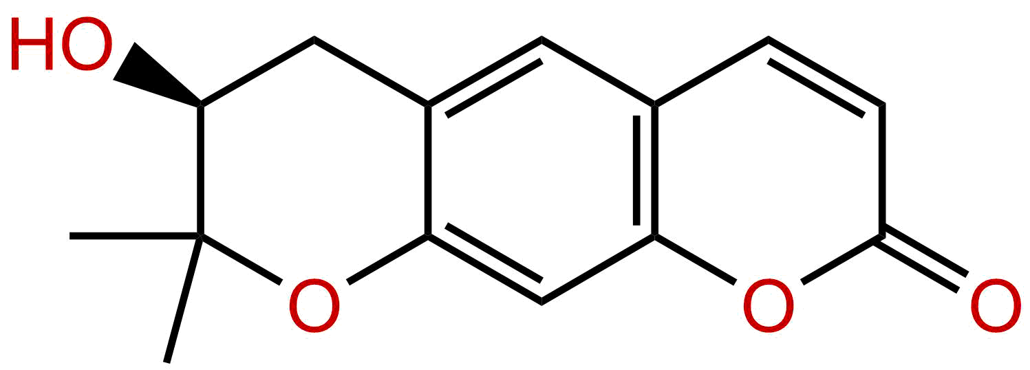
DecursinolCAS No.:23458-02-8
|
||||||||||
 |
|
|
||||||||

| Catalogue No.: | BPF2775 |
| Formula: | C14H14O4 |
| Mol Weight: | 246.262 |
Product name: Decursinol
Synonym name: 3',4'-dihydro-3'-hydroxy-Xanthyletin; Smyrinol; 5993-18-0
Catalogue No.: BPF2775
Cas No.: 23458-02-8
Formula: C14H14O4
Mol Weight: 246.262
Botanical Source:
Physical Description:
Type of Compound: Coumarins
Purity: 95%~99%
Analysis Method: HPLC-DAD or/and HPLC-ELSD
Identification Method: Mass, NMR
Packing: Brown vial or HDPE plastic bottle
Storage: Store in a well closed container, protected from air and light. Put into refrigerate or freeze for long term storage.
Whenever possible, you should prepare and use solutions on the same day. However, if you need to make up stock solutions in advance, we recommend that you store the solution as aliquots in tightly sealed vials at -20℃. Generally, these will be useable for up to two weeks.
The product could be supplied from milligrams to grams
Inquire for bulk scale.
For Reference Standard and R&D, Not for Human Use Directly.
Description:
Decursinol may be a beneficial antimetastatic agent, targeting MMPs and its upstream signaling molecules; it inhibits the proliferation and invasion of CT-26 colon carcinoma cells, might via downregulated ERK and JNK phosphorylation. Aspirin-decursinol has neuroprotective effects, may be closely related to the attenuation of ischemia-induced gliosis and maintenance of antioxidants.
References:
Planta Med. 2013 Nov;79(16):1536-44.
In vitro metabolism of pyranocoumarin isomers decursin and decursinol angelate by liver microsomes from man and rodents.
The aim of this study is to investigate and compare the metabolic rate and profiles of pyranocoumarin isomers decursin and Decursinol angelate using liver microsomes from humans and rodents, and to characterize the major metabolites of decursin and Decursinol angelate in human liver microsomal incubations using LC-MS/MS.
METHODS AND RESULTS:
First, we conducted liver microsomal incubations of decursin and Decursinol angelate in the presence or absence of NADPH. We found that in the absence of NADPH, decursin was efficiently hydrolyzed to Decursinol by hepatic esterase(s), but Decursinol angelate was not. In contrast, formation of Decursinol from Decursinol angelate was mediated mainly by cytochrome P450(s). Second, we measured the metabolic rate of decursin and Decursinol angelate in liver S9 fractions from mice and humans. We found that human liver S9 fractions metabolized both decursin and Decursinol angelate more slowly than those of the mouse. Third, we characterized the major metabolites of decursin and Decursinol angelate from human liver microsomes incubations using HPLC-UV and LC-MS/MS methods and assessed the in vivo metabolites in mouse plasma from a one-dose PK study. Decursin and Decursinol angelate have different metabolite profiles.
CONCLUSIONS:
Nine metabolites of decursin and nine metabolites of Decursinol angelate were identified in human liver microsome incubations besides Decursinol using a hybrid triple quadruple linear ion trap LC-MS/MS system, and many of them were later verified to be also present in plasma samples from rodent PK studies.
PLoS One. 2015 Feb 19;10(2):e0114992.
Single oral dose pharmacokinetics of decursin and decursinol angelate in healthy adult men and women.
The ethanol extract of Angelica gigas Nakai (AGN) root has promising anti-cancer and other bioactivities in rodent models. It is currently believed that the pyranocoumarin isomers decursin (D) and Decursinol angelate (DA) contribute to these activities. We and others have documented that D and DA were rapidly converted to Decursinol (DOH) in rodents. However, our in vitro metabolism studies suggested that D and DA might be metabolized differently in humans.
METHODS AND RESULTS:
To test this hypothesis and address a key question for human translatability of animal model studies of D and DA or AGN extract, we conducted a single oral dose human pharmacokinetic study of D and DA delivered through an AGN-based dietary supplement Cogni.Q (purchased from Quality of Life Labs, Purchase, NY) in twenty healthy subjects, i.e., 10 men and 10 women, each consuming 119 mg D and 77 mg DA from 4 vegicaps. Analyses of plasma samples using UHPLC-MS/MS showed mean time to peak concentration (Tmax) of 2.1, 2.4 and 3.3 h and mean peak concentration (Cmax) of 5.3, 48.1 and 2,480 nmol/L for D, DA and DOH, respectively. The terminal elimination half-life (t1/2) for D and DA was similar (17.4 and 19.3 h) and each was much longer than that of DOH (7.4 h). The mean area under the curve (AUC0-48h) for D, DA and DOH was estimated as 37, 335 and 27,579 h∙nmol/L, respectively. Gender-wise, men absorbed the parent compounds faster and took shorter time to reach DOH peak concentration. The human data supported an extensive conversion of D and DA to DOH, even though they metabolized DA slightly slower than rodents.
CONCLUSIONS:
Therefore, the data generated in rodent models concerning anti-cancer efficacy, safety, tissue distribution and pharmacodynamic biomarkers will likely be relevant for human translation.