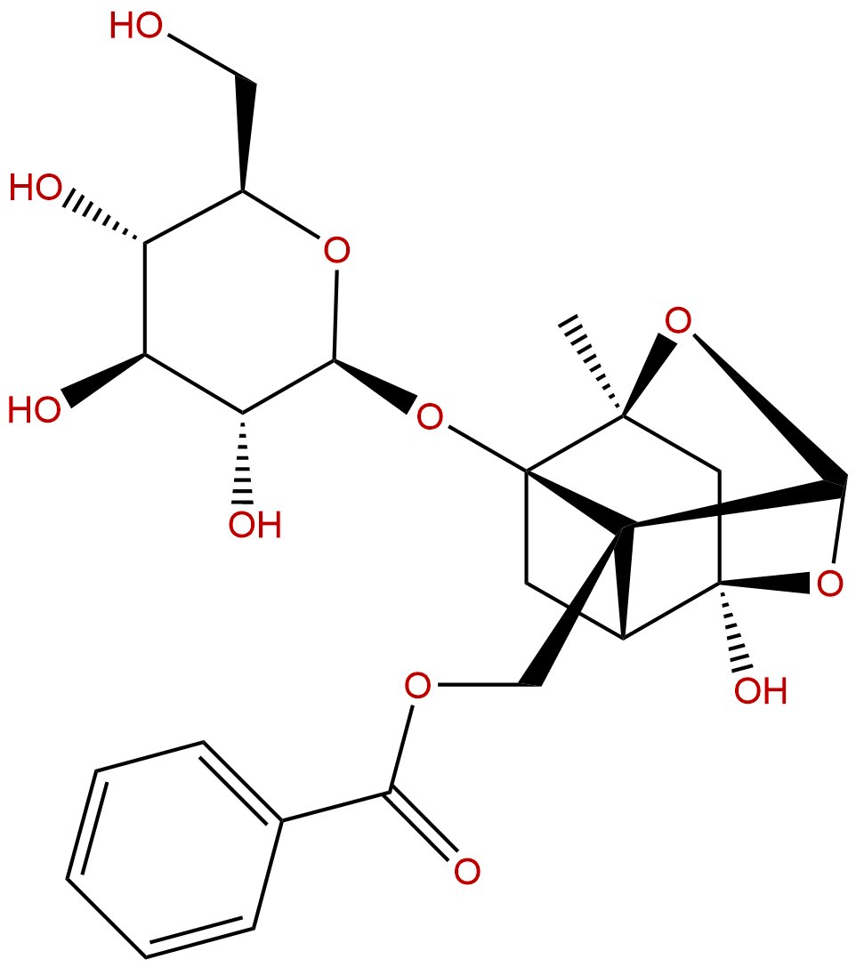
PaeoniflorinCAS No.:23180-57-6
|
||||||||||
 |
|
|
||||||||

| Catalogue No.: | BP1056 |
| Formula: | C23H28O11 |
| Mol Weight: | 480.466 |
Product name: Paeoniflorin
Synonym name: Peoniflorin
Catalogue No.: BP1056
Cas No.: 23180-57-6
Formula: C23H28O11
Mol Weight: 480.466
Botanical Source: Paeonia lactiflora Pall.
Physical Description:
Type of Compound: Monoterpenoids
Purity: 95%~99%
Analysis Method: HPLC-DAD or/and HPLC-ELSD
Identification Method: Mass, NMR
Packing: Brown vial or HDPE plastic bottle
Storage: Store in a well closed container, protected from air and light. Put into refrigerate or freeze for long term storage.
Whenever possible, you should prepare and use solutions on the same day. However, if you need to make up stock solutions in advance, we recommend that you store the solution as aliquots in tightly sealed vials at -20℃. Generally, these will be useable for up to two weeks.
The product could be supplied from milligrams to grams
Inquire for bulk scale.
Description:
Paeoniflorin, a novel heat shock protein-inducing compound, is mediated by the activation of heat shock transcription factor 1 (HSF1), which has antiallergic, anti-obesity, anti-inflammatory and immunoregulatory effects. Paeoniflorin can activate PI3K/Akt signaling pathway to protect the PC12 cell injury induced by Aβ25-35, it protects thymocytes against irradiation-induced cell damage by scavenging ROS and attenuating the activation of the mitogen-activated protein kinases.
References:
Phytomedicine. 2014 Sep 15;21(10):1170-7.
Inhibitory effect of paeoniflorin on methylglyoxal-mediated oxidative stress in osteoblastic MC3T3-E1 cells.
Methylglyoxal (MG) has been suggested to be one major source of intracellular reactive carbonyl compounds. In the present study, the effect of Paeoniflorin on MG-induced cytotoxicity was investigated using osteoblastic MC3T3-E1 cells.
METHODS AND RESULTS:
Osteoblastic MC3T3-E1 cells were pre-incubated with Paeoniflorin before treatment with MG, and markers of oxidative damage and mitochondrial function were examined. Pretreatment of MC3T3-E1 cells with Paeoniflorin prevented the MG-induced cell death and formation of intracellular reactive oxygen species, cardiolipin peroxidation, and protein adduct in osteoblastic MC3T3-E1 cells. In addition, Paeoniflorin increased glutathione level and restored the activity of glyoxalase I to almost the control level. These findings suggest that Paeoniflorin provide a protective action against MG-induced cell damage by reducing oxidative stress and by increasing MG detoxification system. Pretreatment with Paeoniflorin prior to MG exposure reduced MG-induced mitochondrial dysfunction by preventing mitochondrial membrane potential dissipation and adenosine triphosphate loss. Additionally, the nitric oxide and nuclear respiratory factor 1 levels were significantly increased by Paeoniflorin, suggesting that Paeoniflorin may induce mitochondrial biogenesis. Paeoniflorin treatment decreased the levels of proinflammatory cytokines such as TNF-α and IL-6.
CONCLUSIONS:
These findings indicate that Paeoniflorin might exert its therapeutic effects via upregulation of glyoxalase system and mitochondrial function. Taken together, Paeoniflorin may prove to be an
Int Immunopharmacol. 2007 May;7(5):662-73.
Paeoniflorin induced immune tolerance of mesenteric lymph node lymphocytes via enhancing beta 2-adrenergic receptor desensitization in rats with adjuvant arthritis.
Paeoniflorin (Pae), a monoterpene glucoside, is one of the main bioactive components of total glucosides of paeony (TGP) extracted from the root of Paeonia lactiflora. TGP has anti-inflammatory and immunoregulatory effects.
METHODS AND RESULTS:
In this study, we investigated the effects of Pae on inflammatory and immune responses to the mesenteric lymph node (MLN) lymphocytes and the mechanisms by which Pae regulates beta 2-adrenergic receptor (beta 2-AR) signal transduction in adjuvant arthritis (AA) rats. The onset of secondary arthritis in rats appeared around day 14 after injection of Freund's complete adjuvant (FCA). Remarkable secondary inflammatory response and lymphocytes proliferation were observed in AA rats, along with the decrease of anti-inflammatory cytokines interleukin (IL)-4 and transforming growth factor-beta 1 (TGF-beta 1) of MLN lymphocytes, and the increase of pro-inflammatory cytokine IL-2. The administration of Pae (50, 100 mg kg(-1), days 17-24) significantly diminished the secondary hind paw swelling and arthritis scores, reversed the changes of cytokines as discussed above, and further decreased the lowered proliferation of MLN lymphocytes in AA rats. In vitro, Pae restored the previously increased level of cAMP of MLN lymphocytes at the concentrations of 12.5, 62.5 and 312.5 mg l(-1). Meanwhile, Pae increased protein expressions of beta 2-AR and GRK2, and decreased that of beta-arrestin 1, 2 of MLN lymphocytes in AA rats.
CONCLUSIONS:
These results suggested that Pae might induce the Th1 cells immune tolerance, which then shift to Th2, Th3 cells mediated activities to take effect the anti-inflammatory and immunoregulatory effects. The mechanisms of Pae on beta 2-AR desensitization and beta 2-AR-AC-cAMP transmembrane signal transduction of MLN lymphocytes play crucial roles in pathogenesis of this disease.
Planta Med. 1997 Aug;63(4):323-5.
Antihyperglycemic effects of paeoniflorin and 8-debenzoylpaeoniflorin, glucosides from the root of Paeonia lactiflora.
METHODS AND RESULTS:
Paeoniflorin and 8-debenzoylPaeoniflorin were isolated from the dried root of Paeonia lactiflora Pall. (Ranunculaceae). They produced a significant blood sugar lowering effect in streptozotocin-treated rats and had a maximum effect at 25 min after treatment. This hypoglycemic action was also observed in normoglycemic rats only at 1 mg/kg. The antihyperglycemic activity of 8-debenzoylPaeoniflorin seems lower than that of Paeoniflorin. Plasma insulin was not changed in Paeoniflorin-treated normoglycemic rats indicating an insulin-independent action. Also, this glucoside reduced the elevation of blood sugar in glucose challenged rats.
CONCLUSIONS:
Increase of glucose utilization by Paeoniflorin can thus be considered. There are no previous data showing the hypoglycemic activity of Paeoniflorin and/or 8-debenzoylPaeoniflorin in rats.
treatment for diabeteic osteopathy.
HPLC of Paeoniflorin

NMR of Paeoniflorin
